Thursday, May 21, 2020

Saturated Fat: Cutting Through the Noise


The paleo/low-carb world loves to cry about the relative quality of different dietary fats and how they relate to health. They reject the scientific consensus that states that saturated fat (SFA) contributes to cardiovascular disease risk (CVD). Their primary arguments essentially revolve around the notion that we have inadequate data to demonstrate that SFA increases CVD risk. Some will even go so far as to say that SFA is uniquely protective against CVD due to its ability to protect low density lipoprotein particles (LDL) from oxidation. Let's explore these claims.

1) Saturated Fat and Cardiovascular Disease

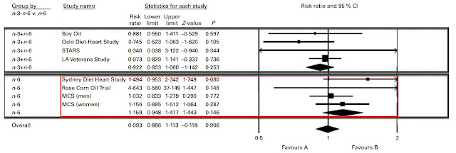
These arguments usually rely heavily on meta-analyses showing no effect of substituting PUFA for SFA on CVD risk in RCTs. But, ultimately these papers are typically just examples of meta-analysis methods being mangled and abused to serve a narrative. Let's look at the typical meta-analyses cited by paleo/low-carb people [1][2][3][4][5]. If we take the time to pick through them it doesn't take long to discover the problem. The methods and inclusion criteria are structured such that highly confounded or otherwise weak studies get lumped in with the high quality studies that are better controlled.

Trials that were unacceptably confounded, like the Sydney Diet Heart Study and the Minnesota Coronary Experiment, manage to sneak their way in. Both of these trials made no delineation between polyunsaturated fatty acids (PUFA) and trans-fats. Other trials with pathetically small amounts of statistical power, like the Rose Corn Oil Trial (n=41 lol), also manage to show up with a shocking amount of weight. If we jam enough weak or poorly controlled trials into our meta-analysis, they will offset the effects observed in the stronger trials and pull our effect toward a null result.


Needless to say, this is ridiculous. The continuous use of these flawed methods creates a sort of Groundhog Day effect that will leave us seeing the same shitty results over and over. Clearly we need better inclusion criteria and better statistical analyses to suss this out. Most meta-analyses aren't actually asking the most pertinent question⁠— can rates of CVD-related mortality be predicted by the degree of LDL-lowering achieved by replacing SFA with PUFA? When we actually investigate the data while taking into account the relevant chain of causality, which is SFA -> LDL -> CVD, we actually see the effect in full swing [6].

Silverman, et al, did a great job of sussing this out. When the data is analyzed in this fashion, we see that reductions in CVD-related mortality are actually perfectly predicted by the degree of LDL-lowering achieved by replacing SFA with PUFA. Using LDL as the moderator variable in the meta-regression, we see that the interventions actually did reduce CVD commensurate with LDL-lowering. Not really a big surprise.


But they also take it a step further and plug the diet trials in alongside a number of other trials using different sorts of LDL-lowering interventions in another meta-regression. We see that the reductions in CVD-related mortality are perfectly predicted by the degree of LDL-lowering relative to other methods of LDL-lowering. The diet trials are exactly where we would expect them to be on the continuum of risk based on the LDL lowering achieved.


Let's look at the trials that met the inclusion criteria in this paper [7][8][9][10]. Notice anything interesting? That's right. These are the trials that typically get cancelled out by weaker trials in the silly-ass meta-analyses that I mentioned earlier.

The results discovered by Silverman, et al, are what we see when we strip away all of the weaker trials that don't belong in the analysis to begin with. This is what happens when we employ a laser-focused inclusion criteria and sound analysis methods. 

Some people might say that the inclusion criteria in this meta-regression is too narrow, and that it is not fair to exclude the other classic fatty acid substitution trials. Interestingly enough, another meta-regression was just published a couple of days ago that includes virtually all of the classic fatty acid substitution trials [11]. Here are the pooled results.


Interesting findings. They are actually perfectly consistent with the guidelines to keep SFA below 10% of calories. We understand that the threshold of effect for SFA is a chronic consumption in excess of approximately 26-30g/day. This is why the guidelines tell us to stay under 10% of calories as SFA, because 10% of a 2000-2500 calorie diet is about 22-27g/day. 10% of calories is just a guideline to keep us under that threshold.

The findings of this second meta-regression also cohere with prospective cohort data when SFA is measured across cohorts as a percentage of calories [12]. Paleo quacks at this point might feel inclined to point out that there are some meta-analyses of prospective cohort studies that find no association between SFA and CVD mortality [13]. But, ultimately it just depends on how we choose to analyze the data, and what data we choose to include.

If we're just pooling median CVD mortality risk between countries based on within-population SFA intake, it's easy to understand how null results are possible. Not all populations are the same. Let me explain.

In America and Europe virtually everyone is eating >30g/day of SFA even in the lowest intake groups [14][15]. In China and Japan virtually everyone is eating <20g/day of SFA even in the highest intake groups [16][17][18]. So of course the relative risk is low or null within or between populations. This is because we're comparing high intakes versus high intakes and low intakes versus low intakes rather than genuinely measuring low versus high.

Take a look at the previous graph. The relative risk of CVD does not seem to increase once one's SFA intake goes beyond 10% of calories (approximately 22-27g/day). So naturally, meta-analyses that are not taking this into consideration will be vulnerable to missing the effect in their results. We need to measure low intakes versus high intakes. Period.

When we dig into the prospective cohort meta-analyses, we see the effect clear as day in cohorts that actually do have a wider range of intake [19][20][21]. We see studies that show intakes ranging from 14-40g/day (Mann, et al) of SFA rather than 30-60g/day of SFA or 7-18g/day of SFA. We see a step-wise increase in CVD-related mortality as a function of SFA intake in those particular cohorts. To hammer this point home, I conducted my own meta-analysis of prospective cohort studies based on my own inclusion/exclusion criteria. You can read more about that here.

But, this is what we see when we are not just comparing high intakes versus high intakes or low intakes versus low intakes and ignoring the threshold effect mentioned above. This is what we see when we are genuinely comparing low intakes versus high intakes in populations that are also crossing the threshold of effect within the range of intake. As a consequence, we see big differences in the relative risk of CVD mortality within those cohorts.

We do also have at least one case of an entire population shifting their average SFA intake from high to low, and seeing massive population-level reductions in CVD-related mortality [22][23]


From 1972-1985, this Finnish population dropped their CVD mortality rates by approximately 30%. The majority of this effect was attributable to the population substituting margarine for butter. Smoking rates did not change much and average BMI actually increased during this time. Back then, the decrease in mortality was attributed to measured reductions in total cholesterol across the population. However, in retrospect we can understand that it was likely the reductions in LDL that followed from ditching the butter.

2) Saturated Fat and LDL Oxidation

At this point we can say with confidence that SFA generally increases CVD risk in humans. But let's explore this idea that SFA uniquely protects LDL from oxidation. As it turns out, this question has actually been investigated from multiple angles. We've actually studied the effect of SFA, monounsaturated fat (MUFA), and PUFA on LDL oxidation [24][25][26]

On balance, there is little to no differential effect of SFA on the lag-time to LDL oxidation when compared to MUFA, and MUFA-rich diets might actually be more protective than SFA-rich diets. While it is true that the PUFA content determines an LDL particle's capacity for potential oxidation, the actual lag-time to oxidation is largely determined by the total antioxidant content of the fat itself [27][28][29][30].


Flax oil, hemp oil, almond oil, safflower oil, sunflower oil, perilla oil, and rapeseed oil are all high-PUFA oils and their effects on LDL oxidation have been studied in detail [31][32][33][34][35][36]. The net effect of these oils is either to have no effect on the rate of LDL oxidation or to decrease the rate of LDL oxidation. It would appear that an LDL particle's susceptibility to oxidation has less to do with PUFA and more to do with antioxidant content of the fats being consumed. 

In addition to increasing the lag time to LDL oxidation, the polyphenols found in these plant-derived oils could also be lowering the total amount of LDL particles through a threefold mechanism, independent of PUFA. Firstly, plant polyphenols may increase hepatic LDL receptor activity in certain cases [37]. Secondly, plant polyphenols may inhibit cholesterol uptake in the gut in certain cases [38]. Thirdly, increasing an LDL particles's resistance to oxidation may increase its binding affinity for LDL receptors and thus lower total LDL [39][40]

This could also provide us with a hint as to why animal fats seem to have such a pronounced effect on both LDL oxidation as well as LDL levels in general [41][42]. Animal fats are as close to isolated fatty acids as you can get in the human diet. Animal fats contain no polyphenols and they have very few antioxidant nutrients, like vitamin E, on average. So, animal fats in general seem to do very little to protect against LDL oxidation or mitigate the LDL-raising effects of SFA.

3) Saturated Fat from Animal Foods

This may seem like specious speculation on my part, however we have actual population-level outcome data supporting the view that animal-derived fats, particularly SFA, are uniquely problematic [43][44][45][46][47]. We also have a lot of data suggesting that many plant sources of SFA may be either neutral or beneficial for LDL and CVD risk, especially when compared to animal-derived SFA [48][49][50][51]. Even coconut oil seems to lower LDL went compared to butter, despite having 30% more SFA by weight.


All together it paints a pretty grim picture for animal-derived fats in general. Suggesting that even if it is the case that SFA uniquely protects LDL from oxidation, it may not be enough to offset the consequences of raising total LDL. Which in turn suggests that even if PUFA increases the rate of LDL oxidation, it doesn't appear to be enough to offset PUFA's LDL-lowering benefits.

In short, if MUFA protects LDL from oxidation to the same degree as SFA (if not better), why prefer SFA at all? SFA seems to offer no unique advantages and is at best a liability for CVD in and of itself. However, it seems that if you must choose a SFA-rich oil, plant-derived oils are very likely superior to animal-derived oils due to how the polyphenols may affect lipid metabolism. It may be true that SFA protects LDL from oxidation. In fact, it is true. But, it is no less true for MUFA, and MUFA might be better at it than SFA. So, why not simply prefer plant-derived MUFA and PUFA overall, and maybe don't sweat the plant-derived SFA in your diet as much.

As an ironic side-note, SFA specifically from cheese seems to have neutral or beneficial effects on LDL as well [52]. I say "ironic" because within the paleo diet's canon, dairy is generally off limits. But, if the paleo/low-carb folks wish to claim a victory in this regard, it would appear that there are also SFA-rich animal foods that don't seem to perturb LDL. But as far as I can tell this effect is strictly related to dairy fat that has not undergone homogenization [53]. This is likely due to the effect of the milk fat globule membrane. This membrane creates a food matrix that appears to mitigate the LDL-raising effects of the SFA. 

4) Linoleic Acid and Cardiovascular Disease

Lastly, there is one additional claim made by paleo/low-carb folks that we should probably investigate as well. The claim that unsaturated oils high in linoleic acid (LA), a plant-derived omega-6 fatty acid, cause CVD. Support for this claim seems to be limited to narrative reviews made by whole-food purists who've somehow managed to get their bullshit published [54]. But let's take a look at the data before we wrap this up.

We already know from what we've discussed above that, generally speaking, SFA restriction leads to reductions in CVD mortality on a population level. But how does LA contribute to CVD risk in isolation? A meta-analysis (with imperfect but albeit better inclusion criteria than most meta-analyses on this topic) on fatty acid substitution RCTs sought to investigate the relationship between LA consumption and coronary heart disease (CHD) mortality and myocardial infarction (MI) [55]


Right off the bat, there are a couple confounded trials we can do away with. Again, the Minnesota Coronary Experiment needs to go. The Finnish Mental Hospital Study also needs to go due to the fact that it wasn't a true RCT and there was potential confounding due to different patient care and differing usages of pharmaceuticals between hospitals. 

Nevertheless, even just eyeballing the damn forest plot it would be obvious that even with the omission of those two trials, the aggregated result would heavily favour PUFA. Just for fun, let's make our own forest plot to see the results.


The results favour replacing SFA with PUFA. The replacement yields an 18% reduction in risk, and is statistically significant (P=0.006). But how might this play out in the real world? These RCTs are investigating fatty acid substitutions by dumping isolated fatty acids on top of existing diets. People eat food. So, can we investigate the effect of LA on a population just eating foods and living their normal lives? 

Epidemiological findings would seem to support the hypothesis that dietary LA confers reductions in CVD mortality [56]. But if you're one of those whacky paleo/low-carb people who absolutely hates nutritional epidemiology due to the supposed unreliability of food frequency questionnaires, you're in luck! Researchers who were aware of this potential issue said screw food frequency questionnaires. They just looked at tissue LA as a direct proxy for intake, and related LA representation in various tissues to CVD mortality [57]. Nice and tidy.


Colour me quirky, but that does not look like an agent of cardiovascular destruction to me. 

One possible rebuttal at this point would be to suggest that this could merely be a reflection of healthy dietary habits, due to the fact that public health and nutrition guidelines have spent decades trumpeting for PUFA. This is known as the "healthy-user bias." It's certainly a plausible explanation. Except that the data seen above includes cohorts from non-English speaking countries that did not have dietary guidelines making any specific recommendations toward fat other than to limit it [58].

So, while the explanation is plausible, it may not actually be very likely. The healthy user bias cannot explain these results. Especially since, at least ostensibly, it would appear that those whose behaviours were at odds with the guidelines (by consuming more dietary fat and not less) actually had better health outcomes if their dietary fats were LA-rich. A truly hilarious amount of mental gymnastics would be required to explain these results.

In conclusion, as a general effect SFA consumption above a threshold of 10% of calories (or 22-27g/day) increases CVD risk in humans through increases in LDL. However, there are SFA-rich foods that can lower LDL and CVD risk due to mitigating factors such as fibre, polyphenols, or unique characteristics of the food matrix itself. It would also appear that dietary LA has an independent beneficial effect on CVD mortality, based on multiple lines of evidence spanning the entire evidence hierarchy. Sorry to say, but this is one situation where the dietary guidelines appear to be correct.

PS. If you like what you've read and want me to continue writing, consider supporting me on Patreon. Every little bit helps! Thank you for reading!

References:

[1] Christopher E Ramsden, et al. n-6 Fatty Acid-Specific and Mixed Polyunsaturate Dietary Interventions Have Different Effects on CHD Risk: A Meta-Analysis of Randomised Controlled Trials. Br J Nutr. 2010 Dec. https://pubmed.ncbi.nlm.nih.gov/21118617/

[2] Christopher E Ramsden, et al. Use of Dietary Linoleic Acid for Secondary Prevention of Coronary Heart Disease and Death: Evaluation of Recovered Data From the Sydney Diet Heart Study and Updated Meta-Analysis. BMJ. 2013 Feb. https://pubmed.ncbi.nlm.nih.gov/23386268/ 

[3] Christopher E Ramsden, et al. Re-evaluation of the Traditional Diet-Heart Hypothesis: Analysis of Recovered Data From Minnesota Coronary Experiment (1968-73). BMJ. 2016 Apr. https://pubmed.ncbi.nlm.nih.gov/27071971/ 

[4] Zoë Harcombe, et al. Evidence From Randomised Controlled Trials Does Not Support Current Dietary Fat Guidelines: A Systematic Review and Meta-Analysis. Open Heart. 2016 Aug. https://pubmed.ncbi.nlm.nih.gov/27547428/

[5] Steven Hamley. The Effect of Replacing Saturated Fat With Mostly n-6 Polyunsaturated Fat on Coronary Heart Disease: A Meta-Analysis of Randomised Controlled Trials.  Nutr J. 2017 May. https://pubmed.ncbi.nlm.nih.gov/28526025/ 

[6] Michael G Silverman, et al. Association Between Lowering LDL-C and Cardiovascular Risk Reduction Among Different Therapeutic Interventions: A Systematic Review and Meta-analysis. JAMA. 2016 Sep. https://pubmed.ncbi.nlm.nih.gov/27673306/

[7] Morris, et al. Controlled Trial of Soya-Bean Oil in Myocardial Infarction. Lancet. 1968 Sep. https://pubmed.ncbi.nlm.nih.gov/4175085/ 

[8] S Dayton, et al. Controlled Trial of a Diet High in Unsaturated Fat for Prevention of Atherosclerotic Complications. Lancet. 1968 Nov. https://pubmed.ncbi.nlm.nih.gov/4176868/ 

[9] P Leren. The Effect of Plasma Cholesterol Lowering Diet in Male Survivors of Myocardial Infarction. A Controlled Clinical Trial. Acta Med Scand Suppl. 1966. https://pubmed.ncbi.nlm.nih.gov/5228820 

[10] Research Committee. Low-fat Diet in Myocardial Infarction: A Controlled Trial. Lancet. 1965 Sep. https://pubmed.ncbi.nlm.nih.gov/4158171/

[11] Lee Hooper, et al. Reduction in saturated fat intake for cardiovascular disease. Cochrane Database of Systematic Reviews. 19 May 2020. https://www.cochranelibrary.com/cdsr/doi/10.1002/14651858.CD011737.pub2/full 

[12] Marianne U Jakobsen, et al. Major Types of Dietary Fat and Risk of Coronary Heart Disease: A Pooled Analysis of 11 Cohort Studies. Am J Clin Nutr. 2009 May. https://pubmed.ncbi.nlm.nih.gov/19211817/

[13] Patty W Siri-Tarino, et al. Meta-analysis of Prospective Cohort Studies Evaluating the Association of Saturated Fat With Cardiovascular Disease. Am J Clin Nutr. 2010 Mar. https://pubmed.ncbi.nlm.nih.gov/20071648/

[14] Margret Leosdottir, et al. Cardiovascular Event Risk in Relation to Dietary Fat Intake in Middle-Aged Individuals: Data From The Malmö Diet and Cancer Study. Eur J Cardiovasc Prev Rehabil. 2007 Oct. https://pubmed.ncbi.nlm.nih.gov/17925631/ 

[15] Kyungwon Oh, et al. Dietary Fat Intake and Risk of Coronary Heart Disease in Women: 20 Years of Follow-Up of the Nurses' Health Study. Am J Epidemiol. 2005 Apr. https://pubmed.ncbi.nlm.nih.gov/15781956/ 

[16] H Y Lee, et al. Serum Fatty Acid, Lipid Profile and Dietary Intake of Hong Kong Chinese Omnivores and Vegetarians. Eur J Clin Nutr. 2000 Oct. https://pubmed.ncbi.nlm.nih.gov/11083485/ 

[17] H Kato, J Tillotson, et al. Epidemiologic Studies of Coronary Heart Disease and Stroke in Japanese Men Living in Japan, Hawaii and California. Am J Epidemiol. 1973 Jun. https://pubmed.ncbi.nlm.nih.gov/4713933/ 

[18] Kenji Wakai, et al. Dietary Intakes of Fat and Total Mortality Among Japanese Populations With a Low Fat Intake: The Japan Collaborative Cohort (JACC) Study. Nutr Metab (Lond). 2014 Mar. https://pubmed.ncbi.nlm.nih.gov/24597664/

[19] J I Mann, et al. Dietary Determinants of Ischaemic Heart Disease in Health Conscious Individuals. Heart. 1997 Nov. https://pubmed.ncbi.nlm.nih.gov/9415002/

[20] Jiaqiong Xu, et al. Dietary Fat Intake and Risk of Coronary Heart Disease: The Strong Heart Study. Am J Clin Nutr. 2006 Oct. https://pubmed.ncbi.nlm.nih.gov/17023718/

[21] A Ascherio, et al. Dietary Fat and Risk of Coronary Heart Disease in Men: Cohort Follow Up Study in the United States. BMJ. 1996 Jul. https://pubmed.ncbi.nlm.nih.gov/8688759/

[22] Erkki Vartiainen, et al. Thirty-five-year Trends in Cardiovascular Risk Factors in Finland. Int J Epidemiol. 2010 Apr. https://pubmed.ncbi.nlm.nih.gov/19959603/

[23] Erkki Vartiainen. The North Karelia Project: Cardiovascular Disease Prevention in Finland. Glob Cardiol Sci Pract. 2018 Jun. https://pubmed.ncbi.nlm.nih.gov/30083543/

[24] P Mata, et al. Effect of Dietary Fat Saturation on LDL Oxidation and Monocyte Adhesion to Human Endothelial Cells in Vitro. Arterioscler Thromb Vasc Biol. 1996 Nov. https://pubmed.ncbi.nlm.nih.gov/8911273/

[25] U S Schwab, et al. The Effect of Quality and Amount of Dietary Fat on the Susceptibility of Low Density Lipoprotein to Oxidation in Subjects With Impaired Glucose Tolerance. Eur J Clin Nutr. 1998 Jun. https://pubmed.ncbi.nlm.nih.gov/9683400/

[26] M J Thomas, et al. Fatty Acid Composition of Low-Density Lipoprotein Influences Its Susceptibility to Autoxidation. Biochemistry. 1994 Feb. https://pubmed.ncbi.nlm.nih.gov/8110785/

[27] Jaume Marrugat, et al. Effects of Differing Phenolic Content in Dietary Olive Oils on Lipids and LDL Oxidation--A Randomized Controlled Trial. Eur J Nutr. 2004 Jun. https://pubmed.ncbi.nlm.nih.gov/15168036/

[28] Evangelia Tsartsou, et al. Network Meta-Analysis of Metabolic Effects of Olive-Oil in Humans Shows the Importance of Olive Oil Consumption With Moderate Polyphenol Levels as Part of the Mediterranean Diet. Front Nutr. 2019 Feb. https://www.ncbi.nlm.nih.gov/pmc/articles/PMC6379345/

[29] Álvaro Hernáez, et al. Olive Oil Polyphenols Decrease LDL Concentrations and LDL Atherogenicity in Men in a Randomized Controlled Trial. J Nutr. 2015 Aug. https://www.ncbi.nlm.nih.gov/pmc/articles/PMC4516770/

[30] Hicham Berrougui, et al. Extra Virgin Olive Oil Polyphenols Promote Cholesterol Efflux and Improve HDL Functionality. Evid Based Complement Alternat Med. 2015 Oct. https://www.ncbi.nlm.nih.gov/pmc/articles/PMC4606102/

[31] Nalini Kaul, et al. A Comparison of Fish Oil, Flaxseed Oil and Hempseed Oil Supplementation on Selected Parameters of Cardiovascular Health in Healthy Volunteers. J Am Coll Nutr. 2008 Feb. https://pubmed.ncbi.nlm.nih.gov/18460481/

[32] Dianne A Hyson, et al. Almonds and Almond Oil Have Similar Effects on Plasma Lipids and LDL Oxidation in Healthy Men and Women. J Nutr. 2002 Apr. https://pubmed.ncbi.nlm.nih.gov/11925464/

[33] Wayne H F Sutherland, et al. Effect of Meals Rich in Heated Olive and Safflower Oils on Oxidation of Postprandial Serum in Healthy Men. Atherosclerosis. 2002 Jan. https://pubmed.ncbi.nlm.nih.gov/11755938/

[34] R Carmena, et al. Effect of Olive and Sunflower Oils on Low Density Lipoprotein Level, Composition, Size, Oxidation and Interaction With Arterial Proteoglycans. Atherosclerosis. 1996 Sep. https://pubmed.ncbi.nlm.nih.gov/8842355/

[35] O Ezaki, et al. Long-term Effects of Dietary Alpha-Linolenic Acid From Perilla Oil on Serum Fatty Acids Composition and on the Risk Factors of Coronary Heart Disease in Japanese Elderly Subjects. J Nutr Sci Vitaminol (Tokyo). 1999 Dec. https://pubmed.ncbi.nlm.nih.gov/10737229/

[36] Nina S Nielsen, et al. Different Effects of Diets Rich in Olive Oil, Rapeseed Oil and Sunflower-Seed Oil on Postprandial Lipid and Lipoprotein Concentrations and on Lipoprotein Oxidation Susceptibility. Br J Nutr. 2002 May. https://pubmed.ncbi.nlm.nih.gov/12010587/

[37] Alberto Dávalos, et al. Red Grape Juice Polyphenols Alter Cholesterol Homeostasis and Increase LDL-receptor Activity in Human Cells in Vitro. J Nutr. 2006 Jul. https://pubmed.ncbi.nlm.nih.gov/16772435/

[38] Shoko Kobayashi. The Effect of Polyphenols on Hypercholesterolemia through Inhibiting the Transport and Expression of Niemann–Pick C1-Like 1. Int J Mol Sci. 2019 Oct. https://www.ncbi.nlm.nih.gov/pmc/articles/PMC6801711/

[39] T J van Berkel, et al. LDL Receptor-Independent and -Dependent Uptake of Lipoproteins. Atherosclerosis. 1995 Dec. https://pubmed.ncbi.nlm.nih.gov/8821464/

[40] G Jürgens, et al. Modification of Human Serum Low Density Lipoprotein by Oxidation--Characterization and Pathophysiological Implications. Chem Phys Lipids. Nov-Dec. https://pubmed.ncbi.nlm.nih.gov/3319231/ 

[41] Ari Palomäki, et al. Effects of Dietary Cold-Pressed Turnip Rapeseed Oil and Butter on Serum Lipids, Oxidized LDL and Arterial Elasticity in Men With Metabolic Syndrome. Lipids Health Dis. 2010 Dec. https://pubmed.ncbi.nlm.nih.gov/21122147/

[42] M Suzukawa, et al. Effects of Fish Oil Fatty Acids on Low Density Lipoprotein Size, Oxidizability, and Uptake by Macrophages. J Lipid Res. 1995 Mar. https://pubmed.ncbi.nlm.nih.gov/7775859/

[43] Marta Guasch-Ferré, et al. Olive oil intake and risk of cardiovascular disease and mortality in the PREDIMED Study. BMC Med. 2014 May. https://www.ncbi.nlm.nih.gov/pmc/articles/PMC4030221/ 

[44] Marta Guasch-Ferré, et al. Associations of Monounsaturated Fatty Acids From Plant and Animal Sources With Total and Cause-Specific Mortality in Two US Prospective Cohort Studies. Circ Res. 2019 Apr. https://pubmed.ncbi.nlm.nih.gov/30689516/

[45] Pan Zhuang, et al. Dietary Fats in Relation to Total and Cause-Specific Mortality in a Prospective Cohort of 521 120 Individuals With 16 Years of Follow-Up. Circ Res. 2019 Mar. https://pubmed.ncbi.nlm.nih.gov/30636521/

[46] Jingjing Jiao, et al. Dietary Fats and Mortality Among Patients With Type 2 Diabetes: Analysis in Two Population Based Cohort Studies. BMJ. 2019 Jul. https://pubmed.ncbi.nlm.nih.gov/31266749/

[47] Geng Zong, et al. Monounsaturated Fats From Plant and Animal Sources in Relation to Risk of Coronary Heart Disease Among US Men and Women. Am J Clin Nutr. 2018 Mar. https://pubmed.ncbi.nlm.nih.gov/29566185/

[48] Lukas Schwingshackl, et al. Effects of Oils and Solid Fats on Blood Lipids: A Systematic Review and Network Meta-Analysis. J Lipid Res. 2018 Sep. https://pubmed.ncbi.nlm.nih.gov/30006369/

[49] C Cox, et al. Effects of Dietary Coconut Oil, Butter and Safflower Oil on Plasma Lipids, Lipoproteins and Lathosterol Levels. Eur J Clin Nutr. 1998 Sep. https://pubmed.ncbi.nlm.nih.gov/9756121/

[50] Chenyan Lv, et al. Effects of dietary palm olein on the cardiovascular risk factors in healthy young adults. Food Nutr Res. 2018 Jul. https://www.ncbi.nlm.nih.gov/pmc/articles/PMC6052506/

[51] O A Tokede and J M Gaziano, L Djoussé. Effects of Cocoa Products/Dark Chocolate on Serum Lipids: A Meta-Analysis. Eur J Clin Nutr. 2011 Aug. https://pubmed.ncbi.nlm.nih.gov/21559039/

[52] Farinaz Raziani, et al. Consumption of regular-fat vs reduced-fat cheese reveals gender-specific changes in LDL particle size - a randomized controlled trial. Nutr Metab (Lond). 2018 Sep. https://www.ncbi.nlm.nih.gov/pmc/articles/PMC6150982/

[53] Fredrik Rosqvist, et al. Potential Role of Milk Fat Globule Membrane in Modulating Plasma Lipoproteins, Gene Expression, and Cholesterol Metabolism in Humans: A Randomized Study. Am J Clin Nutr. 2015 Jul. https://pubmed.ncbi.nlm.nih.gov/26016870/

[54] James J DiNicolantonio and James H O’Keefe. Omega-6 vegetable oils as a driver of coronary heart disease: the oxidized linoleic acid hypothesis. Open Heart. 2018 Sep. https://www.ncbi.nlm.nih.gov/pmc/articles/PMC6196963/

[55] Dariush Mozaffarian, et al. Effects on Coronary Heart Disease of Increasing Polyunsaturated Fat in Place of Saturated Fat: A Systematic Review and Meta-Analysis of Randomized Controlled Trials. PLoS Med. 2010 Mar. https://pubmed.ncbi.nlm.nih.gov/20351774/

[56] Maryam S Farvid, et al. Dietary Linoleic Acid and Risk of Coronary Heart Disease: A Systematic Review and Meta-Analysis of Prospective Cohort Studies. Circulation. 2014 Oct. https://pubmed.ncbi.nlm.nih.gov/25161045/

[57] Matti Marklund, et al. Biomarkers of Dietary Omega-6 Fatty Acids and Incident Cardiovascular Disease and Mortality. Circulation. 2019 May. https://pubmed.ncbi.nlm.nih.gov/30971107/

[58] Wei-Sin Yang, et al. Association Between Plasma N-6 Polyunsaturated Fatty Acids Levels and the Risk of Cardiovascular Disease in a Community-based Cohort Study. Sci Rep. 2019 Dec. https://pubmed.ncbi.nlm.nih.gov/31848413/

Monday, January 27, 2020

Animal Nutrients, Part 4: Vitamin B12


You may have noticed that throughout my Animal/Plant Nutrients series, I don't really go into massive detail about all of the ways these nutrients behave in the human body. I tend to pick an interesting topic specific to each nutrient and weigh in on it. This time I'll be writing about vitamin B12 (B12), and I'll be weighing in on pernicious anemia (PA) specifically.

PA is a relatively common disease, and many of us likely know someone who has been afflicted with it. Both my father and my significant other have PA. My father is currently in his sixties, which is around the age PA tends to set in. My significant other is currently in her late twenties, having developed a rare form of the disease in childhood.

The disease is characterized by a weakened capacity to secrete intrinsic factor (IF) in the stomach [1]. IF binds vitamin B12 and facilitates uptake in the small intestine. As you can imagine, B12 absorption is heavily limited by IF secretion. In those who secrete no IF, only around 1-2% of B12 is absorbed [2]. This means that nutritional doses of B12 have virtually no impact on B12 status in those with the worst IF secretion. Without an appropriate intervention, deficiency is inevitable and the afflicted person will die.

Typically when someone is diagnosed with PA later in life it is assumed they retain at least some IF function and can make use of oral B12 supplements. As such, a simple 1000mcg B12 supplement may be prescribed. If the supplement isn't effective, or becomes less effective with age, the patient may be moved to intramuscular injection therapy (IIT). IIT involves regular appointments with a physician for a single injection of B12 directly into the patient's muscle tissue. This was the therapy that my significant other was placed on from the start when she was first diagnosed with PA as a teenager. For more than a decade she met with a physician once per month for IIT. Eventually it became less and less effective, which necessitated her self-injecting at home.

Over time she told me more about her history with the disease and how it was currently affecting her. Naturally I started researching it like crazy. I found many papers detailing the effectiveness of IIT, and I was left with the distinct impression that this was the only viable treatment method. Toward the end of my digging I used Twitter to ask Chris Masterjohn about the plausibility of using creatine to spare B12 in the methylation cycle. My idea was that she could perhaps extend the effectiveness of each injection. While he acknowledged that the mechanism was plausible, he also explained that it was probably pointless. He further explained that high dose oral supplementation was likely just as effective as IIT.

I fired up PubMed and immediately started looking for anything I could find relating to sublingual (SL) B12 supplementation as an alternative treatment for PA. Indeed, I found a number of papers detailing the effectiveness of SL B12 [3][4][5][6]. I remember immediately becoming irate. I can understand the unfortunate reality of regular self-injecting for type I diabetics, because there is no viable alternative for them. But I could not understand this. Why was the standard of care for PA so draconian when there were other demonstrably viable alternatives? It didn't make sense to me.

Shortly after my discovery, I suggested to my significant other that she start on 5000mcg of SL B12 per day. I advised her to continue with IIT if the SL B12 proved ineffective. Sure enough, however, from the moment she started SL B12 almost a year ago, she has not needed a single injection and presents with no symptoms of B12 deficiency. She reports that the SL B12 works better than IIT acutely, and is overall more effective on a month-to-month basis as well. Only time will tell whether or not the effect will wane, but I suspect it won't.

So, why does SL B12 work so well? The short answer is that B12 molecules are small enough to passively diffuse through tissues and into your circulation. So, when you hold 5000mcg of B12 under your tongue, it has to go somewhere. As previously mentioned, even without IF 1-2% of oral B12 is absorbed through passive diffusion in the gut. Which means that nutritional doses of B12 do virtually nothing for PA, but it also means that mega-doses can probably do a lot. For this reason, I highly suspect that if one finds IIT more effective than SL B12, it is merely because the SL dose was not high enough. I find it difficult to believe that dosing 20mg or more of SL B12 wouldn't overcome the issue.

Just last summer, approximately four months after my partner began SL B12, a trial was run wherein IIT was compared head-to-head against SL B12 [7]. Guess what. The effectiveness of SL B12 was equal, if not superior, to IIT. It overcomes the literal pain-in-the-ass of IIT, and probably has some other unique advantages as well. My only hope is that the standard of care adjusts in order to accommodate this effective, less invasive method of treating patients with PA. It seems as though I'm not alone with my hopes, either [8][9].

PS. If you like what you've read and want me to continue writing, consider supporting me on Patreon. Every little bit helps! Thank you for reading!

References:

[1] Bizzaro and Antico. Diagnosis and classification of pernicious anemia. Autoimmun Rev. 2014 Apr. https://www.ncbi.nlm.nih.gov/pubmed/24424200

[2] Berlin H, et al. Oral treatment of pernicious anemia with high doses of vitamin B12 without intrinsic factor. Acta Med Scand. 1968 Oct. https://www.ncbi.nlm.nih.gov/pubmed/5751528

[3] Bolaman Z, et al. Oral versus intramuscular cobalamin treatment in megaloblastic anemia: a single-center, prospective, randomized, open-label study. Clin Ther. 2003 Dec. https://www.ncbi.nlm.nih.gov/pubmed/14749150

[4] Delpre G, et al. Sublingual therapy for cobalamin deficiency as an alternative to oral and parenteral cobalamin supplementation. Lancet. 1999 Aug. https://www.ncbi.nlm.nih.gov/pubmed/10475189

[5] E Nyholm, et al. Oral vitamin B12 can change our practice. Postgrad Med J. 2003 Apr. https://www.ncbi.nlm.nih.gov/pmc/articles/PMC1742688/

[6] Andrès E, et al. Effects of oral crystalline cyanocobalamin 1000 μg/d in the treatment of pernicious anemia: An open-label, prospective study in Ten Patients. Curr Ther Res Clin Exp. 2005 Jan. https://www.ncbi.nlm.nih.gov/pubmed/24672108

[7] Bensky MJ, et al. Comparison of sublingual vs. intramuscular administration of vitamin B12 for the treatment of patients with vitamin B12 deficiency. Drug Deliv Transl Res. 2019 Jun. https://www.ncbi.nlm.nih.gov/pubmed/30632091

[8] Josep Vidal-Alaball, et al. Oral vitamin B12 versus intramuscular vitamin B12 for vitamin B12 deficiency. Cochrane Database Syst Rev. 2005 Jul. https://www.ncbi.nlm.nih.gov/pmc/articles/PMC5112015/

[9] Catherine Qiu Hua Chan, et al. Oral Vitamin B12 Replacement for the Treatment of Pernicious Anemia. Front Med (Lausanne). 2016 Aug. https://www.ncbi.nlm.nih.gov/pmc/articles/PMC4993789/

Friday, January 3, 2020

Measuring Nutrient Density: Calories vs Weight


The Nutrient Density Cheat Sheet considers the nutritional yield of foods per standard serving size. Serving size is a food measurement based on the weight of a given food, and is meant to reflect the food portion sizes that people typically eat. Recently the Nutrient Density Cheat Sheet was criticized by a somewhat fanatical vegan. She claimed that by considering nutrition per serving, I was unfairly biasing all of my scores toward animal foods. She doubled down and further claimed that if my scores were recalculated using calories instead of weight, leafy green vegetables would certainly be revealed as the most nutrient dense foods on the list.

We went back and forth about the correct methodology for some time. It took quite a while for her to coherently distill her objections down into something I could actually work with. Essentially, she asserts that water and fibre confound the weight and volume measurements in unacceptable ways, and that calories are a more accurate way to measure nutrient density. She suggested that I completely remove weight as a variable, and do my calculations strictly with nutrients per calorie. So, I decided to humour her and I did precisely what she asked me to do.

I spent a couple hours going back and recalculating all of the nutrient data by calories instead of serving weight. The results are still adjusted for bioavailability, nutrient absorption capacity, and metabolic conversion inefficiencies (my methods for each adjustment are detailed in an earlier blog post here). There are absolutely no weight measurements considered in the nutrition per calorie scoring calculations. Here are the results:


Animal foods seem to still come out on top. However, in order to insulate myself against criticisms regarding my nutrient yield adjustments, I also produced two other unadjusted scores. They use the same methodology— one is nutrient density per serving, and the other is nutrient density per calorie. Neither is adjusted for nutrient bioavailability, nutrient absorption capacity, or metabolic conversion inefficiencies. It's literally just each nutrient divided by its respective DRI and divided by calories. Results are summed across all nutrients per food, and the results across all foods are sorted and ranked. Here are those results:


As you can see some leafy green vegetables do get a boost, but ultimately animal foods are still dominating the top of the list. But, why is this? Leafy greens are low calorie and animal foods are higher calorie. So, it seems intuitive that leafy greens would be some of the lowest calorie foods, so why does measuring nutrient density per calorie actually produce these counter-intuitive results? It's because dividing nutrition by calories just gives you a silly little ratio. That's it. The results don't actually have to favour low calorie foods at all. The results just favour foods that have a similar ratio of nutrition to calories, which can include both high and low calorie foods.

Highly nutritious, high calorie foods (like Atlantic salmon) get similar scores as poorly nutritious, low calorie foods (like spinach). Think about it. If you divide 1000 by 100, you get 10. If you divide 10 by 1, you get 10. It's just a ratio. It tells you nothing about realistic portion sizes or caloric density. Which is why dividing nutrition by calories is a foolish and uninformative way of quantifying nutrient density. For example, black coffee can be found in both the adjusted and unadjusted scores when calculating nutrition per calorie. Which could leave one with the false impression that black coffee is a great source of nutrients, or is at least comparable to oysters or mussels. However, one would have to consume approximately 1.7 litres in order to exceed the RDA of a single essential nutrient found in coffee (riboflavin in this case). Whereas eating just 10g of either mussels or oysters yields more than the RDA of vitamin B12. 10g is barely the size of the tip of your finger. Whereas 1.7 litres is an insane amount of coffee to drink to get the RDA of riboflavin.

One of this vegan's primary arguments in support of nutrition per calorie measurements was that humans have a limited calorie budget (approximately 2000 kcal/day), so assessing nutrition per calorie is best. While it's true we all eat within a similar calorie budget, it's not true that measuring nutrition per calorie actually gives you much meaningful insight into the calorie yield of a food. It's just a ratio. The foods on the top of the list need not be low calorie foods at all.

Ultimately, my position is that calories are a subjective value-judgement. Calories are something you assess completely independently of nutrient density, and you increase or decrease calories according to your goals. On this basis alone, I suggest that factoring calories into the nutrient density score necessarily injects subjective bias into the results. Nutrition divided by calories has a number of unacceptable drawbacks. 

Firstly, considering nutrition per calorie assumes that calories are always a disadvantage. Secondly, it punishes foods for having essential nutrition. Both essential amino acids and essential fatty acids contain calories, so they actually lower the nutrient density score. Which clearly doesn't make any sense. Lastly, it just doesn't actually give you any meaningful information about calories. So, why even bother? Nutrition per serving also has significant interpretive challenges, but they are far less severe, and far less limiting, than measuring nutrition per calorie. Serving size simply gives you a better approximation of how humans interact with food, and that's what matters.

PS. If you like what you've read and want me to continue writing, consider supporting me on Patreon. Every little bit helps! Thank you for reading!

Monday, December 9, 2019

Nutritional Ketosis and Muscle Hypertrophy


A commonly misunderstood concept in the low carb world is the balance between muscle anabolism and muscle catabolism. The ketogenic diet (KD) has the capacity to perturb this balance negatively, though it is not guaranteed. This is because different macronutrients affect both lean body mass (LBM) and fat mass (FM) differently when considered in isolation. Let me explain.

  • Protein is both anabolic and sparing to LBM, but catabolic to FM.
  • Carbs are sparing of LBM and FM, but anabolic to neither.
  • Fat is catabolic to LBM, but anabolic to FM.

This may appear to be a bit of an oversimplification, but in complete isolation this is essentially how the macronutrients behave under eucaloric conditions. The capacity for anabolism and catabolism will vary as we vary these macronutrients in proportion to each other in the diet. We would expect a high carb, high protein diet to maximally spare LBM. We'd also expect a high protein, KD to spare LBM, but perhaps not as effectively due to the lack of carbohydrate.

When we enter into nutritional ketosis, we deplete liver glycogen and must synthesize glucose by breaking down protein and liberating amino acids (AA). This can be protein in the diet or protein on our body. Eventually we can use other substrates like glycerol and aldehydes to synthesize glucose, but the contribution of AAs to gluconeogenesis (GNG) will always be substantial. This is why we sometimes hear low carb advocates claim that carbs are "non-essential". This is because when we don't eat them, we synthesize them.

However, we cannot rely entirely on dietary protein to satisfy our body's entire demand for glucose. For example, if our acute need for glucose exceeds our capacity to digest, absorb, and metabolize AAs from our diet to glucose, we will be pulling those amino acids from our skeletal muscle instead [1]. This seems to be true in the fed and fasted state while in nutritional ketosis.

Even if we could satisfy 100% of our glucose requirements with dietary AAs in the fed state, we still have to sleep at some point. During sleep, we're fasting by definition and relying on endogenous AAs to synthesize glucose. When protein and calories are matched between a KD and a non-ketogenic diet (nKD), a nKD will typically be more sparing of LBM [2][3]. On a nKD, we can rely on liver glycogen to maintain euglycemia throughout the night, and should awake with only modest ketones in the morning. This added benefit of carbohydrate is part of what serves to maximally spare LBM. The more we can rely on our liver glycogen to supply our body with glucose, the less we have to rely on AAs from skeletal muscle catabolism.

All this being said, it is certainly possible to gain muscle on a KD, despite the catabolic stimulus being very strong. It is likely that we merely need to provide adequate protein and a sufficiently robust anabolic stimulus, like resistance training [4]. It is likely that protein needs are going to be higher on a KD in order to achieve the same balance between anabolism and catabolism that can be achieved on a nKD. However, a KD will always cost us anabolic potential even if we do experience net increases in LBM. This means that even if we made gains, we probably could have made better gains (or at least we could have made the same gains with less effort) on a nKD. 

Ultimately, either we're spending dietary AAs on glucose instead of spending them to build muscle, or we're breaking down already built muscle by liberating AAs to spend on glucose. Glucose isn't free. Either way we lose anabolic potential.

Key points:
  • Muscle hypertrophy occurs when anabolism outweighs catabolism. 
  • We have an obligate need to catabolize lean tissue while in ketosis.
  • Ketogenic diets unavoidably cost us anabolic potential by default.
  • Amino acids used to make glucose cannot be used to build muscle.
  • Typical gains are still achievable in ketosis, but require extra protein.

PS. If you like what you've read and want me to continue writing, consider supporting me on Patreon. Every little bit helps! Thank you for reading!

References:

[1] Claire Fromentin, et al. Dietary Proteins Contribute Little to Glucose Production, Even Under Optimal Gluconeogenic Conditions in Healthy Humans. Diabetes. May 2013. https://www.ncbi.nlm.nih.gov/pmc/articles/PMC3636601

[2] Greene, DA, et al. A Low-Carbohydrate Ketogenic Diet Reduces Body Mass Without Compromising Performance in Powerlifting and Olympic Weightlifting Athletes. J Strength Cond Res. December 2018.
https://www.ncbi.nlm.nih.gov/pubmed/30335720

[3] Wood, RJ, et al. Preservation of fat-free mass after two distinct weight loss diets with and without progressive resistance exercise. Metab Syndr Relat Disord. June 2012.
https://www.ncbi.nlm.nih.gov/pubmed/22283635

[4] Antonio Paoli, et al. Ketogenic Diet and Skeletal Muscle Hypertrophy: A Frenemy Relationship? J Hum Kinet. August 2019.
https://www.ncbi.nlm.nih.gov/pmc/articles/PMC6724590

Wednesday, November 27, 2019

Why We Grow What We Grow


Chris Kresser recently appeared as a guest on the Joe Rogan Experience to discuss and challenge the credibility of the recent film, The Game Changers. During this interview, Kresser had made the statement that staple crops like corn and soy are actually nutrient poor, and that perhaps we should be moving away from our current model of industrial agriculture and focusing on a system that provided more nutrient dense foods.

We can debate the sustainability of different agricultural systems at a different time. For now I just want to discuss his claims regarding the nutritional content of our staple crops. I actually shared this sentiment about our staple crops before I created the Nutrient Density Cheat Sheet, a nutrition ranking tool that uses minimally biased metrics to score and compare over 500 different common foods. After my work on that project was done, it was absolutely clear to me why we choose the staple crops that we choose. It's because they're fucking badass, and for no other reason. It is absolutely insane just how nutrient dense these foods are before we process and refine them.

According to Wikipedia, the most popular grain crops in the world are: corn, rice, wheat, and soy. Kresser claims that these foods are nutrient poor and don't make a very meaningful contribution to the diet. So, what does Kresser recommend instead? When answering this question, Kresser often references the work of Mat Lalonde, and has used Mat's work to defend organ meats, herbs and spices, nuts and seeds, and cocoa as being among the most nutrient dense foods available. But, is this actually true? Are these foods actually the most nutrient dense options we can find? It depends on how we look at it.

Let's break it down. Firstly, Mat's scale only considers essential nutrition per 100g of food, and does not include essential amino acids or essential fatty acids. The scale also does not adjust for bioavailability, nutrient absorption capacity, or nutrient conversion inefficiencies. Needless to say, it is very incomplete. However, as it turns out, my scale does take those things into consideration, or at least it tries to approximate those things based on the best available literature. The Cheat Sheet also includes essential amino acids and essential fatty acids. Lastly, it considers the nutrient density per serving, because serving size better reflects how we interact with different foods. Here are the results when we use my scale to compare our staple crops to Kresser's top picks:


When considered in their whole food state, our staple crops are actually incredibly nutrient dense per serving and can likely contribute a great deal to our diet. Some of Kresser's picks are great, and others are not so good when considering nutrient density per serving. Because he cited food categories instead of individual foods, I was generous and actually took the best food from within each category he listed instead of taking an average. That way we're only looking at the champions of each group.

As we can see, organs are incredibly nutrient dense and can make an incredible addition to the diet. I've written about that before. Herbs and spices are actually pretty nutrient poor per serving, coming in dead last out of everything on the list. They're really not anything to write home about. Nuts and seeds are great, but actually not as good as wheat or soy. Cocoa isn't that great by comparison, either. Keep in mind that I'm considering our staple crops in the cooked state, as well. These numbers aren't confounded by things like dehydration. If we take an average of the total nutrition of each group, we see that our staple crops have an average score of 66, whereas Kresser's picks have an average score of 50. 

I guess the take-home message is, don't make claims before you're in full possession of the relevant facts. While it actually is true that Kresser's top selection of nutrient dense foods actually ranks higher when we consider nutrition per 100g, it is also true that nobody actually eats 100g of dried clove. Virtually nobody eats 100g of nuts or cocoa either. When we consider these foods as people typically consume them, it's obvious why we choose the staple crops that we choose. Provided we're considering these foods in their whole state, they're awesome. 

PS. If you like what you've read and want me to continue writing, consider supporting me on Patreon. Every little bit helps! Thank you for reading!

Wednesday, October 16, 2019

Sugar Doesn't Cause Diabetes, and Ketosis Doesn't Reverse It


One of the only clinical diagnostic criteria for type II diabetes mellitus (T2DM) is a persistently elevated blood glucose (BG) level. Chasing down the cause of this BG elevation has been the subject of an enormous investigation for many decades. Many researchers disagree about the ultimate pathogenesis and pathophysiology of the disease state, and in the 21st century we have the fortune of being able to see these disagreements play out in real time online. Actual PhDs doing actual pivotal research have taken to using social media platforms like Twitter and publicly accessible academic platforms like Research Gate to debate the merits and consistency of the available data. In these places you will find an enormous breadth of opinions— some plausible, some preposterous, and some that surely reflect reality quite accurately.

Some people argue that sugar causes the disease. Others blame fat and animal products. Some even say it is merely an inescapable consequence of aging. People have many conflicting opinions about T2DM. I'm not a researcher. I'm not an expert. But for what it's worth, I'd like to share my thoughts on this subject. To do this, I want to use a bit of a different format with this post. I'm going to state my position in point form, and then I'm going to defend it by breaking each point down. So let's get to it.
  1. Fatty acids cause insulin resistance, but glucose shows it to you.
  2. Diabetes is a disease of excess calories.
  3. Carbohydrate restriction is therapeutic, not curative.
  4. Ketosis is functional diabetes, therefore also therapeutic.
Before we dive into dissecting each one of these points, we need a little crash course in cellular physiology. It won't be painful, I promise. We're just going to briefly review the Randle cycle. 

The Randle cycle is basically a series of biochemical pathways inside of your cells that signal which fuel substrates are to be burned. In one arrangement, a cell will preferentially burn glucose. In the other arrangement, a cell will preferentially burn fatty acids (FA). Under normal conditions, the arrangement is dependent on the relative proportions of each available substrate. For example, after a carbohydrate rich meal, the cycle will favour burning carbohydrates. After a fat rich meal, the cycle will favour burning FAs. Here we see a diagram of these mechanisms in action. 


On the left and right we see glucose and long-chain fatty acids (LCFA) being transported into the cytosol of an adipocyte. Here we see how the availability of either substrate dictates which substrate is to be burned as fuel. Let's start with glucose.

When glucose enters through glucose transporter type 4 (GLUT4), it is driven through an enzymatic pathway known as glycolysis and cleaved in half. This gives us two three-carbon units of pyruvate, which enter the mitochondria. Not seen here, one pyruvate goes on to participate in the Citric Acid (TCA) cycle, and the other pyruvate gets converted to a two-carbon unit called Acetyl-CoA by the pyruvate dehydrogenase complex (PDH). The extra carbon is lost as CO2. If there is too much pyruvate, this drives up Acetyl-CoA and inhibts PDH. This is a form of negative feedback. When the PDH complex is inhibited by Acetyl-CoA, excess pyruvate is shunted out of the cell as lactate.

When LCFA enter the cell, they're broken apart into two-carbon units called long chain fatty Acyl-CoA (LC-FAC) by long chain fatty Acyl-CoA synthetase (LC-FACS). These enter the mitochondria through carnitine palmitoyltransferase 1 (CPT1). From there, each LC-FAC is converted to Acetyl-CoA through beta-oxidation (β-ox). Notice how excess Acetyl-CoA does not have a negative feedback system inside the mitochondria. Instead, excess Acetyl-CoA drives up mitochondrial citrate, and thus drives up cytosolic citrate. Cytosolic citrate is the signal to inhibit GLUT4 and phosphofructokinase 1 (PFK-1). This means that if enough citrate is generated, glucose is both inhibited from entering the cell and inhibited from entering glycolysis.

Further downstream we discover that FAs do in fact have their own negative feedback system. The consequence of continuing to drive up cytosolic citrate is to eventually produce Malonyl-CoA. Which stimulates fatty acid synthase (FAS) for the purposes of storing excess fatty acids through the synthesis of new triglycerides (TG). Malonyl-CoA also inhibits CPT-1, preventing LC-FAC from being transported into the mitochondria.

There we go. We now understand the basics of the Randle cycle. Now let's talk about how this is fundamentally relevant for understanding my position on T2DM.

1. Fatty acids cause insulin resistance, but glucose shows it to you.

Let's refer back to the graphic above. We know that FAs in the cell uniquely inhibit GLUT4. It all depends on the relative proportions of FAs to glucose that are available to the cell. The more FAs you have in the cell, the more resistant that cell will be to translocating GLUT4 to the cell surface to take up glucose. Thus, the activity of the PDH complex will be impaired as the cell begins to preferentially burn FAs as fuel [1]. This impairs oxidative glucose disposal and increases non-oxidative glucose disposal [2]. Essentially, when you eat a high-fat diet, you lose glycogen in your liver, your cells will make the switch to FA-burning, and oxidative disposal of glucose will go down. When you finally do get a bolus of glucose, it will serve to replete glycogen stores and will be disposed of through non-oxidative pathways. More astute individuals in the low-carb community will recognize this whole situation as "physiological insulin resistance", which they rightly state is a non-pathological state.

However, since merely increasing the availability of FAs to a cell will powerfully reduce its sensitivity to insulin [3], we can surmise that the relative insulin sensitivity of a cell is dictated by its FA burden. As such, if one's cells are overburdened with FAs, glucose concentrations in the blood will be higher due to the Randle cycle's effects on insulin sensitivity and glucose disposal [4]. When you challenge the body with glucose while the body is attempting to burn through a surplus of FAs, glucose is going to have to wait outside the cell in the blood until those FAs are burned. When we see this happen in T2DM, it is very tempting to blame the carbs in the diet as being the ultimate cause of the persistently elevated glucose we see in the blood. But the glucose isn't actually to blame. The elevated BG is merely reflecting the competition between energy substrates based on the relative availability of each substrate to the body's cells, particularly the adipocytes. When there is too much fat, glucose cannot be burned.

Am I attempting to imply that a high-fat diet causes T2DM? No, not at all. But the effects of high-fat feeding on glucose metabolism gives us insight into the roles of carbs and fats in T2DM. Which leads me to my next point.

2. Diabetes is a disease of excess calories.

We can confidently say that the symptoms of T2DM are made uniquely worse by carbohydrates, as removing them has profound utility with regards to symptom management [5]. Nobody can really dispute this credibly at this point. However, there is a massive amount of evidence indicating that the primary source of insulin resistance in T2DM at the cellular level is the FA burden of the actual cell [6]. So, what determines the FA burden of a cell? By and large, the answer is every Taubesian acolyte's least favourite word—calories. Calories, calories, calories. 

But isn't insulin a fat storage hormone? Aren't the carbohydrates responsible for sequestering fat into our fat tissue? Yes and no. Insulin stores fat and displaces lipolysis proportionate to the energy provided by the carbohydrates that stimulated the insulin. So yes, if you were somehow able to learn the history of each TG in your adipose tissue, you'd discover that the vast majority of those TGs originated as FAs consumed in the diet. However, insulin wasn't really required to store any of them. If we refer back to the diagram of the Randle cycle for a moment, we discover that merely overburdening an adipocyte with FAs will stimulate TG synthesis. Whether those TGs were stored in the adipocyte through insulin or stored passively by merely consuming fat, it is the total calorie balance of the diet that determines the TG content of the adipocyte at the end of the day [7].

There is a growing body of evidence that suggests that sufficient caloric restriction leads to the remission and perhaps the reversal of T2DM in a manner that is entirely predicted by fat mass loss [8][9]. Fat mass loss is dictated by caloric intake. No more, no less. 


So, to recap. If you overeat carbohydrates, you will indeed store every ounce of fat you consume. However, calorie for calorie, if you overconsume fat, you will drive up TG synthesis and store every ounce of excess fat you consume as well. At the end of the day, total calorie intake minus your total energy expenditure determines fat loss or fat gain. If you overeat enough calories chronically, eventually your adipocytes become so overloaded with FAs that they're indefinitely stuck in the FA-burning position. It is similar to only being able to turn one handle on a faucet, when you should be able to turn both.

3. Carbohydrate restriction is therapeutic, not curative.

Let's go back to BG for a moment. Why is carbohydrate restriction beneficial for T2DM? If it's the fat inside the cell that's causing the problem, why should there be benefits to restricting carbohydrates at all? The short answer is that consuming excess fat carries fewer short-term negative symptoms when compared to glucose in the context of T2DM. When we consume excess fat, it may be readily stored in a number of places. Even though some of these storage depots are problematic, like the liver or even the blood, fat stored in these places causes very few noticeable symptoms under certain conditions. Don't get me wrong, though. It's still not a good thing to store ectopic fat, or to store fat in the blood. However, when you consume glucose while adipocytes and visceral fat stores are experiencing an overload of fat (as with T2DM), all hell breaks loose.

The body does not prefer to store glucose as fat [10]. It never has, and it never will. Glucose that can't be burned is shunted out of cells as lactate. Once the lactate is in the blood it goes to the liver and participates in the Cori cycle to be recycled into glucose. It is then sent back into the blood [11]. Typically, lactate is returned to the liver to be converted to glucose to replete hepatic glycogen. The body knows that while you're burning FAs as fuel, the liver needs extra glucose. But T2DM typically presents with a scenario wherein both subcutaneous fat stores as well as hepatic fat stores are maxed. This impairs the liver's ability to store glycogen. The liver and the adipose are both paddles in a game of Pong, with glucose and lactate as the ball. This is confirmed by the higher levels of plasma and urinary lactate associated with T2DM [12]. As one would expect, this effect can be observed in people with certain glycogen storage disorders as well [13]. In T2DM, this merry-go-round repeats itself indefinitely, until the FA burden on the adipocytes can be resolved. 

But, at this point you might be wondering why this merry-go-round is damaging. Personally, I don't actually think the glucose itself is damaging, but what inevitably results from that glucose is damaging. In this state, you cannot have a glut of BG without a glut of blood lactate, and that is problematic. Lactate can cause anything from hypoxia to tissue necrosis, and pretty much any of the comorbidities associated with T2DM. In fact, some people with T2DM have experienced acute lactic acidosis from taking metformin, a drug that inhibits the Cori cycle [14]. Inhibiting the Cori cycle increases lactate levels in the blood [15][16]. I'm not trying to scare diabetics out of taking metformin. Not at all. In fact, one of the reasons metformin is beneficial is because it opens the loop. If the lactate can't go to the liver to be converted to glucose, it may be redirected to the kidney to be excreted in the urine. This is because the liver and the kidney are two of the body's largest lactate disposal sites. If you take the liver out of the equation, you may very well urinate the excess lactate more easily, or even metabolize it in the kidney more easily. 

I suspect that a combination of metformin and a low-carbohydrate diet could be a powerful tool for reducing many of the comorbidities associated with T2DM. In my estimation, the lactate probably has more to do with these comorbidities than does the glucose itself. But removing the glucose removes the lactate. So, removing the glucose has tremendous utility in T2DM symptom management. Until the FA burden on the adipocytes can be rectified, one cannot expect to be able to tolerate a glucose challenge. It is for this reason that I'm willing to say that the reduction in diabetic symptoms associated with carbohydrate-restriction may very well constitute T2DM remission. However, this is not T2DM reversal. Reversal would imply that one has restored glucose tolerance. Mechanistically, the only durable path toward genuine T2DM reversal would be rigorous calorie restriction, regardless of the macronutrient ratio of the diet. Though, a low carbohydrate diet would likely be less rocky along the way. 

Additionally, as a consequence of the body not preferring to store glucose as fat, you have an innate preference to dispose of glucose through oxidative pathways. This generates a lot of metabolites and byproducts of metabolism that are inherently damaging, and even more damaging in the context of T2DM. Substances like methylglyoxal, reactive oxygen species, and other breakdown products. These products disrupt metabolism and can damage a number of tissues. They can also further disrupt insulin signalling through oxidative damage of the insulin receptor substrate proteins. When you swap the carbohydrates for fat in the diet, there is a greater opportunity to safely store the energy you've consumed. Because the body can safely store this energy, and there is no pressing need to burn it immediately, there is considerably greater leeway with carbohydrate-restrictive diets when we're considering T2DM and its associated comorbidities. 

4. Ketosis is functional diabetes, therefore also therapeutic.

This statement may seem provocative and hyperbolic, but I'm absolutely serious. If you compare ketotic metabolism to diabetic metabolism, there are some spooky similarities.


Pretty much all of the hallmarks of T2DM overlap with the hallmarks of nutritional ketosis, with the exception of glucose, insulin, and lactic acid. This is why ketosis is therapeutic for T2DM. Ketosis removes the primary source of the bulk of the comorbidities associated with T2DM, but the underlying pathology still remains. The insulin resistance persists. The high energy status, and many of its long-term consequences, likely persists as well. In short, ketosis is T2DM without the hyperglycemia. Ketosis takes a dysfunctional state, and makes it more functional by removing the challenge to the glucose disposal system while it is impaired. It doesn't reverse the dysfunctional state. It merely improves the situation by masking some of the associated symptoms.

PS. If you like what you've read and want me to continue writing, consider supporting me on Patreon. Every little bit helps! Thank you for reading!


References:

[1] Chokkalingam K., et al. High-fat/low-carbohydrate diet reduces insulin-stimulated carbohydrate oxidation but stimulates nonoxidative glucose disposal in humans: An important role for skeletal muscle pyruvate dehydrogenase kinase 4. J Clin Endocrinol Metab. January 2007. https://www.ncbi.nlm.nih.gov/pubmed/17062764

[2] Bisschop P.H., et al. Dietary fat content alters insulin-mediated glucose metabolism in healthy men. Am J Clin Nutr. March 2001. https://www.ncbi.nlm.nih.gov/pubmed/11237931

[3] Stephen F. Burns, Sheryl F. Kelsey, and Silva A. Arslanian. Effects of an Intravenous Lipid Challenge and Free Fatty Acid Elevation on In Vivo Insulin Sensitivity in African American Versus Caucasian Adolescents. Diabetes Care. February 2009. https://www.ncbi.nlm.nih.gov/pmc/articles/PMC2628707

[4] Siôn A. Parry, Rachel M. Woods, Leanne Hodson, and Carl J. Hulston. A Single Day of Excessive Dietary Fat Intake Reduces Whole-Body Insulin Sensitivity: The Metabolic Consequence of Binge Eating. Nutrients. August 2017. https://www.ncbi.nlm.nih.gov/pmc/articles/PMC5579612/

[5] Sarah J. Hallberg, et al. Effectiveness and Safety of a Novel Care Model for the Management of Type 2 Diabetes at 1 Year: An Open-Label, Non-Randomized, Controlled Study. Diabetes Ther. April 2018. https://www.ncbi.nlm.nih.gov/pmc/articles/PMC6104272/

[6] Taylor R., et al. Remission of Human Type 2 Diabetes Requires Decrease in Liver and Pancreas Fat Content but Is Dependent upon Capacity for β Cell Recovery. Cell Metab. October 2018. https://www.ncbi.nlm.nih.gov/pubmed/30078554

[7] Kevin D. Hall and Juen Guo. Obesity Energetics: Body Weight Regulation and the Effects of Diet Composition. Gastroenterology. May 2017. https://www.ncbi.nlm.nih.gov/pmc/articles/PMC5568065/

[8] Lean M.E., et al. Primary care-led weight management for remission of type 2 diabetes (DiRECT): an open-label, cluster-randomised trial. Lancet. February 2018. https://www.ncbi.nlm.nih.gov/pubmed/29221645

[9] Roy Taylor. Calorie restriction for long-term remission of type 2 diabetes. Clin Med (Lond). January 2019. https://www.ncbi.nlm.nih.gov/pmc/articles/PMC6399621/

[10] Acheson K.J., Schutz Y., Bessard T., Anantharaman K., Flatt J.P., Jéquier E. Glycogen storage capacity and de novo lipogenesis during massive carbohydrate overfeeding in man. Am J Clin Nutr. August 1988. https://www.ncbi.nlm.nih.gov/pubmed/3165600

[11] Chen Y.D., Varasteh B.B., and Reaven G.M. Plasma lactate concentration in obesity and type 2 diabetes. Diabetes Metab. August 1993. https://www.ncbi.nlm.nih.gov/pubmed/8293860

[12] Jean L. J. M. Scheijen, et al. L(+) and D(−) Lactate Are Increased in Plasma and Urine Samples of Type 2 Diabetes as Measured by a Simultaneous Quantification of L(+) and D(−) Lactate by Reversed-Phase Liquid Chromatography Tandem Mass Spectrometry. Exp Diabetes Res. March 2012. https://www.ncbi.nlm.nih.gov/pmc/articles/PMC3310144/

[13] Hagen T., Korson M.S., and Wolfsdorf J.I. Urinary lactate excretion to monitor the efficacy of treatment of type I glycogen storage disease. Mol Genet Metab. July 2000. https://www.ncbi.nlm.nih.gov/pubmed/10924273

[14] E. Fitzgerald. Metformin associated lactic acidosis. BMJ. September 2009. https://www.bmj.com/content/339/bmj.b3660

[15] Huang W., Castelino R.L., and Peterson G.M. Lactate Levels with Chronic Metformin Use: A Narrative Review. Clin Drug Investig. November 2017. https://www.ncbi.nlm.nih.gov/pubmed/28836132

[16] DeFronzo R., Fleming G.A., Chen K., and Bicsak T.A. Metformin-associated lactic acidosis: Current perspectives on causes and risk. Metabolism. February 2016. https://www.ncbi.nlm.nih.gov/pubmed/26773926